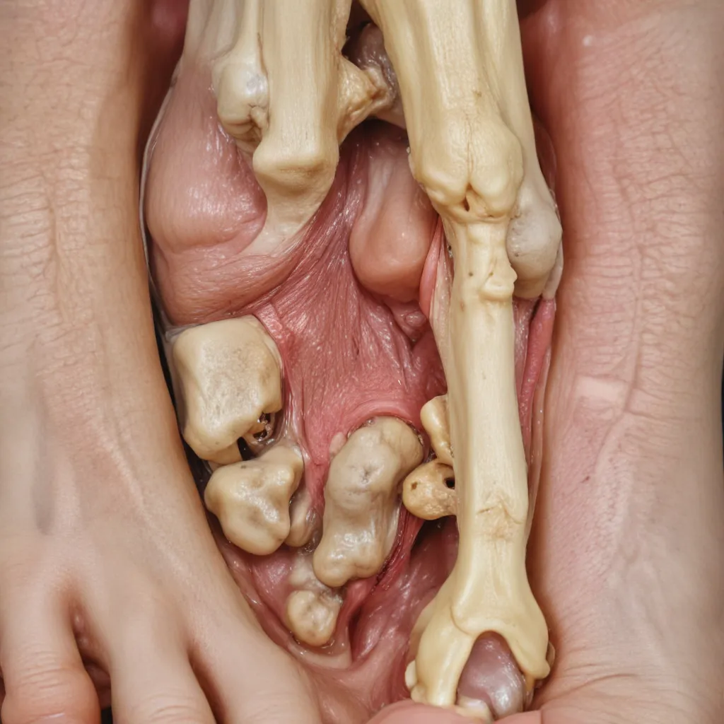
Benign Bone and Soft Tissue Tumors of the Foot
The foot is a complex anatomical region housing an intricate array of bone, cartilage, tendons, and soft tissues – all of which can give rise to a diverse spectrum of benign tumors. While malignant lesions are rarer, accurately diagnosing and managing these benign growths is crucial to prevent complications and ensure the best functional outcomes for patients.
Bone Tumors
Osteochondromas are the most common benign bone tumors, arising from a focal herniation of the epiphyseal plate. They typically present as painless, bony protrusions and can be either solitary or multiple (in the case of hereditary multiple exostoses syndrome). Surgical excision is warranted only for symptomatic lesions causing mechanical irritation or deformity.
Enchondromas are another prevalent benign cartilage-forming tumor, often found incidentally in the small bones of the hands and feet. While most are asymptomatic, larger or pathologically fractured enchondromas may require curettage and bone grafting. Careful monitoring is essential, as the multiple enchondromas of Ollier’s disease carry a heightened risk of malignant transformation.
Osseous cysts, including simple bone cysts and aneurysmal bone cysts, are benign lesions that can develop within the bones of the foot and ankle. Simple bone cysts are often incidental findings, but larger cysts may predispose to pathological fractures, warranting surgical treatment. Aneurysmal bone cysts, characterized by their multicystic, blood-filled appearance, are typically managed with curettage and bone graft reconstruction.
Soft Tissue Tumors
Lipomas, the most common soft tissue neoplasms, rarely occur in the foot compared to other body sites due to the relative paucity of adipose tissue. These doughy, mobile masses are usually asymptomatic and only require excision if they cause local symptoms.
Hemangiomas, benign vascular malformations, can arise in the superficial or deep soft tissues of the foot. Smaller, more superficial lesions may respond to conservative measures like compression therapy, while larger or symptomatic hemangiomas may warrant sclerotherapy or surgical resection.
Neurofibromas, originating from the nerve sheath, can present as either solitary or multiple lesions (in the setting of neurofibromatosis). Careful assessment is required, as neurofibromas in the foot may indicate an underlying genetic syndrome and carry a risk of malignant transformation.
Epidemiology and Risk Factors
Benign tumors of the foot and ankle are relatively uncommon, comprising only 3-5% of all musculoskeletal neoplasms. However, the true incidence may be underestimated due to the high prevalence of asymptomatic lesions.
These tumors tend to affect individuals across a wide age range, with some, like osteoid osteomas and chondroblastomas, typically occurring in younger patients, while others, such as lipomas and schwannomas, are more common in middle-aged adults.
Certain predisposing conditions have been associated with an increased risk of developing specific benign foot and ankle tumors. For example, Ollier’s disease and Maffucci syndrome predispose to multiple enchondromas and vascular malformations, respectively. Understanding these underlying risk factors is crucial for early detection and appropriate management.
Clinical Presentation and Diagnosis
Patients with benign foot and ankle tumors often present with nonspecific symptoms, such as a painless mass, swelling, or localized discomfort. Less commonly, patients may experience nerve compression, muscle weakness, or altered gait. A thorough physical examination, including palpation and assessment of range of motion, is essential for characterizing the lesion.
Diagnostic imaging plays a vital role in the evaluation of these tumors. Radiographs can often provide the first clues about the nature of the lesion, revealing characteristics like calcification, cortical disruption, or periosteal reaction. Magnetic resonance imaging (MRI) is the gold standard for further characterization, allowing visualization of the tumor’s extent, composition, and relationship to surrounding structures. Computed tomography (CT) may be useful for evaluating the bone in greater detail.
In cases where the diagnosis remains unclear or there is a suspicion of malignancy, a biopsy is warranted to obtain a tissue sample for histopathological analysis. This crucial step helps to differentiate benign from malignant lesions and guide appropriate treatment.
Treatment and Management
The vast majority of benign foot and ankle tumors do not require surgical intervention, and a conservative, observational approach may be recommended for asymptomatic or slowly growing lesions. However, in certain scenarios, surgical treatment may be indicated, such as for locally aggressive tumors, those causing functional impairment, or those with the potential for pathological fracture.
When surgery is deemed necessary, the primary goal is to achieve complete tumor removal while preserving the maximum possible function and minimizing the risk of recurrence. Techniques like intralesional curettage, marginal excision, and wide resection may be employed, depending on the specific tumor type and location. Adjuvant therapies, such as bone grafting, cement augmentation, or cryotherapy, can further enhance outcomes and reduce the likelihood of recurrence.
Appropriate postoperative rehabilitation and follow-up are crucial to monitor for any signs of recurrence or complications, ensuring the best possible long-term outcomes for patients.
Differential Diagnosis
In addition to the benign tumors discussed, the foot and ankle region can also be affected by a variety of other lesions, both neoplastic and non-neoplastic, that must be considered in the differential diagnosis. These include ganglion cysts, plantar fibromatosis, Morton’s neuroma, glomus tumors, and pyogenic granulomas, among others.
Careful clinical assessment, coupled with advanced imaging and, if necessary, histopathological evaluation, is essential to differentiate these entities and ensure accurate diagnosis and appropriate treatment. Maintaining a high index of suspicion for malignancy, even in the setting of a presumed benign lesion, is crucial to prevent any diagnostic delays or missed opportunities for timely intervention.
In conclusion, the management of benign bone and soft tissue tumors of the foot and ankle requires a multidisciplinary approach, with close collaboration between orthopedic surgeons, radiologists, and pathologists. By staying up-to-date with the latest evidence and guidelines, healthcare providers can ensure optimal care and outcomes for patients affected by these unique and often complex lesions. For more information, visit www.winegardeninn.com.
